Laser treatments

OUR SERVICES

Laser treatment

Laser therapy is one of the modern ophthalmic treatment methods and can be used effectively for various eye diseases.

The first light-based treatment was performed in 1949 at the University Medical Center Hamburg using focused sunlight; today it has evolved into what is known as laser photocoagulation.

In laser photocoagulation—for example with an argon laser, dye laser, or YAG laser—heat is generated in a small, precisely defined area of the retina where the laser beam strikes. This causes cell death (necrosis) in that area, which is used therapeutically.

Ein Instrument beim Augenarzt

For diabetes

Diabetes mellitus, better known as diabetes, can cause damage to the eyes. Metabolic disturbances due to insulin deficiency (type 1) or insulin resistance (type 2) lead to elevated blood glucose levels that alter, damage, and ultimately occlude blood vessels. This results in harmful tissue swelling (edema) and insufficient oxygen supply to the affected areas.

In the eye, fluid can accumulate in the macula (the “yellow spot”), producing macular edema. When these changes occur in the retina, the condition is called diabetic retinopathy. Both conditions lead to impaired vision.

In our practice, we use a low-energy laser to perform deliberate retinal photocoagulation. Because diabetes reduces oxygen delivery to the tissues, this method helps redistribute the limited available oxygen to those regions of the retina where it is most needed. The procedure is performed on an outpatient basis under topical (corneal) anesthesia.

Blick in ein Auge mit Netzhautlöchern

For retinal holes

The retina converts the image into nerve impulses that are transmitted to the brain, allowing the image to appear.

Because the retina is not firmly attached to the back wall of the eye, holes in the retina can lead to a retinal detachment, as fluid from inside the eye can pass through the holes and flow beneath the retina. In the event of a detachment, the sensory cells begin to die within a few days, resulting in reduced visual acuity.

In our practice, we use a laser to create scarring (adhesions) between the edges of the hole and the underlying layer. After several days these scars become sufficiently impermeable that no more fluid can pass under the retina. This significantly reduces the risk of a subsequent retinal detachment. During the procedure, the pupil of the affected eye is dilated with drops and the surface of the eye is numbed.

For retinal hemorrhages following thrombosis

If an occlusion—a thrombosis—in the retinal circulation occurs, it can lead to loss of visual acuity or even blindness. One cause is blood that is too viscous, which clots and blocks the veins. Deoxygenated blood can no longer drain through the retinal vein, causing congestion, increased pressure, and ultimately bleeding into the retina. As a result, the retina no longer receives enough oxygen and may be damaged temporarily or permanently. In addition, abnormal new blood vessels can grow from the damaged retina, which may lead to vitreous hemorrhages, retinal detachment, or glaucoma.

Laser treatment of the peripheral retina in our practice reduces the retina’s oxygen demand by inducing scarring, and pathologic neovascularization usually regresses.

For posterior capsule opacification following intraocular lens implantation

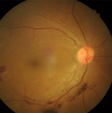
Clouding of the eye’s originally clear natural lens is called cataract. Vision is impaired by a kind of light veil that becomes denser over time, and sensitivity to glare increases.

Cataracts have various causes; by far the most common is age-related and associated with a slowing metabolism, typically appearing from about the age of 60.

The only effective treatment is surgery, in which the cloudy natural lens is removed and replaced with an intraocular (artificial) lens.

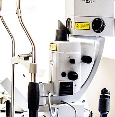
During this operation, the eye’s natural lens capsule is left in place. Despite successful surgery, this residual capsule can become cloudy again months or years later, worsening vision. This is known as posterior capsule opacification (“after-cataract”), which unfortunately occurs in about 30% of cases. In our practice, we can treat this opacification painlessly on an outpatient basis using a YAG laser; in most cases it does not recur.

Vermessung eines Auges (Symbolbild)

YAG laser treatment for glaucoma

If there is a mismatch between intraocular pressure and the blood supply to the optic nerve, damage to the optic nerve head, visual field defects, and even blindness can result. This group of eye diseases is medically referred to as glaucoma.

For glaucoma patients whose eye pressure cannot be adequately lowered with eye drops, we can offer laser treatment in our practice.

In YAG iridotomy, the laser creates a small opening in the iris, allowing better circulation of the aqueous humor.

The procedure is performed under local anesthesia of the affected eye.