Dry eye

OUR SERVICES

Dry eyes / office eye syndrome

One in five patients who consult an ophthalmologist in Germany suffers from dry eyes—a disorder of the natural tear film. These symptoms manifest themselves as burning, itching, or tearing.

These symptoms are collectively referred to as office eye syndrome, as they occur particularly when we are sitting at a computer or otherwise concentrating and forget to blink.

However, various diseases such as diabetes, rheumatism, or thyroid dysfunction can also cause insufficient tear production.

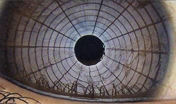
In our practice, we use various tests to determine the extent of the tear film disorder. The Schirmer test is important here: small strips of absorbent paper are placed in the conjunctival sac to measure the time it takes for them to become saturated. By applying a dye to the surface of the eye and asking you not to blink, we can determine how stable the tear film is.

In addition to special eye drops, small changes in everyday behavior can often help, e.g., taking breaks when working at a computer screen, drinking enough fluids, or improving the supply of fresh air.

Dry eyes should be taken seriously, because what may seem like a minor annoyance at first can lead to disease of the eye surface and thus to a loss of vision.

Eine ältere Frau mit Office Eye Syndrom reibt sich das Auge
OUR SERVICES

SBM O.S.A. - Ocular Surface Analyzer Dry Eye Analyzer

Diagnostic platform for the classification of dry eye

Dry eye is a complex, very common condition that is extremely troublesome for patients. Meibomian gland dysfunction (MGD) is the most common cause of dry eye. Morphological changes in the glandular tissue can be visualized using O.S.A. as a digital infrared image. The length and width of the meibomian glands are recorded and the extent and area of failure of the meibomian glands are indicated as a percentage.

The O.S.A. “Dry Eye” platform offers the following examinations in one device:

  • Interferometry: Classification of the lipid layer for assessing Meibomian gland function (before and after IPL treatment)
  • Meibography: Quantification of the Meibomian glands.
    Measurement of the tear meniscus: Measurement of the tear meniscus at up to 5 points to quantify the tear film.
  • Auto Nibut measurement: The tear breakup time is automatically determined using a grid projected onto the cornea. This allows the stability of the mucin layer and the entire tear film to be assessed.
  • Blepharitis (Demodex) test: In addition to Meibomian gland dysfunction, a common cause of dry eye symptoms

Your ophthalmologist can use the O.S.A. to quickly determine what type of dry eye disease you have and thus recommend further treatment.

The O.S.A. measurement results provide answers to the following questions:

  • Disturbed lipid layer due to Meibomian gland dysfunction?
  • Pathologically reduced tear production?
  • Blepharitis?
  • Is IPL the appropriate treatment for my patient?

The extensive measurements are carried out by the assistant staff. Based on the results, your ophthalmologist can make an informed recommendation for dry eye treatment.

eyemed Augenärzte - OSA-Gerät

Auto-meibography is used to visualize and evaluate morphological changes in the glandular tissue as a digital infrared image. For targeted treatment of meibomian gland dysfunction using E-Eye technology to stabilize the tear film, IDRA supports the course and success of the prescribed therapy.

Meibom-Drüsen-Erkennung

AUTO interferometry:

Auto-interferometry can be used to visualize the stability and thickness of the lipid layer before and after IRPL treatment with the E-Eye.

AUTO-NIBUT

The tear film break-up time can be used to assess the overall stability of the tear film between blinks. The key advantage is that this measurement is completely non-invasive and does not involve staining the tear film with fluorescein. (Non-invasive break-up time)

Auto-Nibut

Tear meniscus height

With different magnifications, you can measure the height of the tear meniscus and assess its characteristics along the lower eyelid margin. All results are automatically converted into a standardized grading scale.

Classification of the degree of redness

The redness of the conjunctiva is automatically and objectively documented. The vessels of the bulbar and limbal conjunctiva are evaluated and their degree of redness is compared with the classification sheets.

Rötungsgrad des Auges

Pupillometry

Measurement of pupil response to light with and without glare: scotopic, mesopic, photopic.

Pupillometrie

Tear film analysis

Individual analysis of the tear film enables rapid, detailed, and structural examination of tear composition. Analysis of all layers: lipid layer, aqueous layer, mucin layer.

Additional features:

White-to-white measurement, blepharitis (demodex), yellow filter for fluorescence examination