Ophthalmological examination

OUR SERVICES

Eye examinations and treatments for the front and back sections of the eye

In addition to standard examinations to determine visual acuity, in combination with eyeglass lens consultation (including progressive lenses) and eyeglass prescriptions, as well as contact lens consultations and check-ups, we offer a wide range of special eye examinations and treatments for the front and back of the eye using state-of-the-art diagnostics and effective therapy options, as well as other service-oriented services related to the eye.

Messung der Sehstärke bei einer Patientin

Macular and retinal diseases:

Fluorescent angiography

Imaging of the vessels at the back of the eye using a fluorescent dye (fluorescein) is called fluorescein angiography (FAG). It is an important diagnostic tool for various retinal and macular diseases, such as diabetes mellitus (diabetic retinopathy, diabetic macular disease), vascular occlusions, neovascularization, age-related macular degeneration (AMD), retinal inflammation, and tumors.

To perform the angiography, the pupil must be dilated with eye drops. Fluorescein is a dye and not a contrast agent. The dye reaches the vessels of the eye via the blood circulation after about 20 seconds. Digital single and video images are then taken and evaluated.

OCT

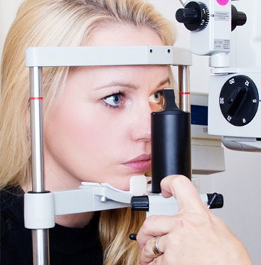
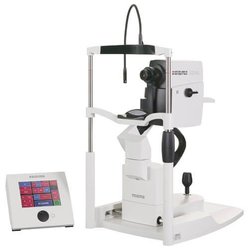
OCT is an abbreviation for optical coherence tomography. With the help of a harmless laser scanner, high-resolution cross-sectional images of the back of the eye can be taken.

With OCT, we have the ability to produce high-resolution retinal layer images of the macula in our practice. This non-invasive method is now the gold standard in the diagnosis of macular and retinal diseases such as holes in the center of the retina (macular holes), the presence of a thin scar tissue on the spot of sharpest vision (macula), also known as epiretinal gliosis, sugar changes in the retina (diabetic retinopathy, diabetic maculopathy), vascular occlusions, and, of course, age-related macular degeneration (AMD). Nerve fiber layer thicknesses can also be measured, which is helpful in the diagnosis of glaucoma.

The procedure is non-contact, painless, and safe.

Untersuchungsgerät Spectralis OCT

Fundus photography

The fundus camera takes precise photographs of the back of the patient’s eye. This is done to document the findings, in particular of the retina, optic nerve, and blood vessels, in high quality. It is used to track the progression of diseases, for diagnosis, and to document the findings.